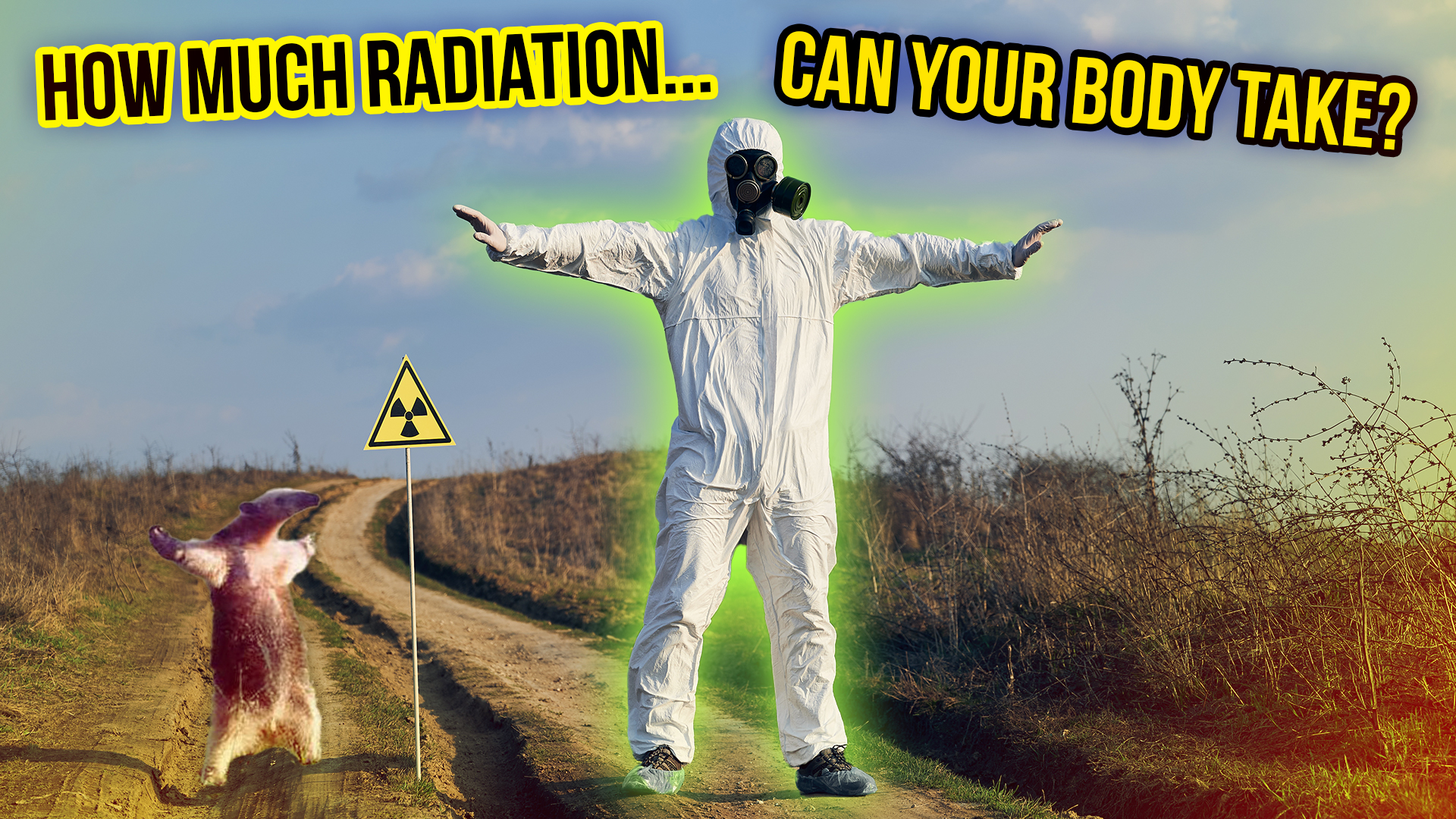
How Much Radiation Can the Human Body Take and are Dental X-Rays Actually Safe
 If you regularly visit the dentist, chances are you have sat at least once for a set of dental x-rays. The procedure is usually straightforward: first, the dentist will drape a protective lead vest over your chest and abdomen, then place x-ray film in a plastic holder in your mouth and the x-ray machine against the side of your head. Finally, they will retreat behind a shielded wall to activate the machine, the actual exposure typically taking no more than one second. But as you are sitting there, waiting for the process to end, several questions may have crossed your mind. Why, if the x-rays are dangerous enough to warrant a lead vest, is your head left unshielded? And if a simple lead vest is enough to block the x-rays, why does the dentist need to hide behind a shielded wall? How powerful are the x-rays anyway? How dangerous are they? And just how much radiation can the human body absorb without risking cancer – or worse? By now you are probably tired of me telling you that the answer to these questions is “it’s complicated,” but alas: it’s complicated.
In order to answer the question “how much radiation is too much radiation?”, we must first define what exactly we mean by “radiation.” In physics, the term “radiation” typically refers to electromagnetic radiation – that is, forms of energy which propagate by means of electric and magnetic fields. Electromagnetic radiation lies on a spectrum, with lower-energy, longer-wavelength microwaves on one end, and higher-energy, shorter-wavelength Gamma rays on the other. The visible spectrum – that is, the electromagnetic wavelengths which are detectable by the human eye – lie somewhere in the middle, between 380 and 750 nanometers. For our purposes, however, we are interested only in so-called ionizing radiation – that is, electromagnetic waves with enough energy to ionize or strip electrons off atoms and damage the DNA of living cells. This includes all the wavelengths past the visible spectrum – that is, ultraviolet rays, X-rays, and Gamma Rays, with the boundary between non-ionizing and ionizing radiation typically being defined as 124 nanometers. Thus, despite the claims of certain conspiracy theorists, our cell phones and microwave ovens cannot give us cancer as these devices operate at energies too low to produce serious genetic damage. Still, there are plenty of other reasons not to stick your head in a microwave.
Another source of ionizing radiation is from high-energy subatomic particles, the three types most commonly encountered being alpha particles, beta particles, and neutrons. Both alpha and beta particles are produced by the natural decay of radioactive elements like Uranium, the former being composed of two protons and two neutrons and the latter being identical to electrons. Neutrons, while occasionally produced via the spontaneous fission of radioactive elements, are typically encountered as a byproduct of nuclear fission chain reactions in reactors and nuclear weapons. Occasionally, other kinds of ionizing particles like protons are also encountered at densities and energies high enough to damage living tissue – for example, in scientific or medical particle accelerators.
But regardless of the type of ionizing radiation encountered, the mechanism of action is the same: the radiation damages the DNA within the exposed cells, resulting in one of two outcomes. In most cases, the cell detects the genetic damage and either repairs it or undergoes a process known as apoptosis or “cell suicide,” preventing the damaged DNA from being replicated. Thanks to these defence mechanisms the body can safely absorb mild doses of radiation such as those produced by cosmic rays from space, radioactive elements in the earth, and medical x-rays. However, if the dose is large enough and enough cells are damaged at once, a nasty chain of symptoms known collectively as Acute Radiation Syndrome or ARS may result. First observed in survivors of the atomic bombings of Hiroshima and Nagasaki, ARS typically first presents as severe nausea, vomiting, and abdominal pain. Skin directly exposed to radiation may initially redden like a sunburn and later swell up, depending on how deeply the radiation has penetrated into the tissue. From here the symptoms only get worse, and may include weakness, fatigue, headache, bloody vomit and stools, hair loss, and cataracts – the latter three resulting from severe damage to the cells of the digestive tract, hair follicles, and lenses of the eye. However, around 24 hours following exposure some victims may seem to spontaneously recover, their symptoms appearing to disappear for several days. This is known as the “walking ghost” phase, and results from certain tissues being more sensitive to radiation than others. Cells undergoing rapid division – such as those of the stomach, sexual organs, and bone marrow – are the most sensitive – hence why one of the first symptoms of ARS is nausea and vomiting. Massive exposure to radiation causes the bone marrow to stop producing blood cells, resulting in a condition known as aplastic anemia. This not only cripples the immune system, leaving the body vulnerable to infection, but also significantly hinders the healing of burns, cuts, and other injuries. However, the cells already in the blood – which are less affected by radiation – remain in circulation for around a month, and thus the immune system initially continues to operate normally. After a few days, however, these cells become depleted, and the victim’s condition rapidly begins to deteriorate. As organs shut down, blood vessels collapse, and tissues literally liquefy, the victim will experience delirium, coma, excruciating pain which even the most powerful opioids cannot touch, and, inevitably, death.
But even relatively mild doses of radiation can still have serious health effects. While in the vast majority of cases the combination of natural genetic repair mechanisms and apoptosis prevents cells from propagating radiation-damaged DNA, once in a while a cell’s DNA becomes mutated in such a way that it starts dividing and multiplying out of control – a condition better known as cancer. And since rapidly-dividing sex cells are particularly sensitive to radiation damage, exposure of the gonads or a developing embryo to even moderate amounts of radiation can result in birth defects.
But exactly how much radiation constitutes a mild, moderate, or severe dose? Well, this is where things gets complicated, for radiation exposure is a stochastic process, meaning it is highly probabilistic and there is no linear, predictable relationship between radiation exposure and negative health effects. Thus, while it is generally the case that a higher radiation dose will result in more severe health effects, there is no way of predicting the precise effects a particular dose will have on a particular person. This is true of all carcinogenic processes. For example, while smoking more cigarettes certainly increases one’s risk of cancer, it is on one hand entirely possible for someone to smoke a pack a day for 80 years and never develop cancer – or, on the other hand, to smoke a single cigarette and immediately develop cancer. It is even possible that mild doses of radiation may actually be beneficial by stimulating the body’s DNA repair mechanisms – an effect known as radiation hormesis – and for more on that, please check out our previous video The Curious Case of the Widespread Radioactive Apartments. Yet despite the complex, unpredictable nature of radiation exposure, health physicists have nonetheless developed a complex system of units, measures, and recommended doses in an attempt to keep workers in nuclear power plants, research laboratories, medical imaging departments, and other high-radiation environments safe and healthy. So buckle up and take a deep breath, because yes: there will be math involved.
Radiation exposure is typically measured in terms of energy absorbed by a given volume or mass of matter. The first standard unit for radiation exposure was the Röntgen, adopted in 1928. Named after German physicist Wilhelm Röntgen, the discoverer of X-rays, one Röntgen was initially defined as the amount of ionizing radiation capable of inducing one statcoulomb of electric charge in one cubic centimetre of dry air. But while an important milestone in quantifying radiation exposure, the Röntgen was only applicable to air and not other materials like human tissue. Thus, in 1945 American medical physicist Herbert Parker introduced the Röntgen Equivalent Physical or Rep, equivalent to 93 ergs of energy absorbed by one gram of matter. In 1953 the Rep was replaced by a new unit known as the Rad. Initially defined in the centimetre-gram-second or CGS system as 100 ergs of energy absorbed by one gram of matter, the Rad is now defined in the Systeme Internationale or SI system as 0.01 Joules of energy per kilogram of matter. The Rad itself was subsequently replaced in 1975 by a new SI unit known as the Gray. Named after British nuclear physics pioneer Louis Robert Gray, the Gray is defined as one Joule of energy absorbed by one kilogram of matter. While the Gray is the de facto standard unit of radiation absorption in much of the world, certain laboratories and industries – especially in the United States – still continue to use the Rad and even sometimes the Röntgen.
However, both the Rad and the Gray measure radiation absorption in any kind of material – be it air, water, plastic, concrete, or anything else – and do not account for the particular effects of radiation on human tissue. As mentioned before, certain tissues are more sensitive than others to radiation, with the gonads, stomach, and bone marrow being among the most sensitive and the brain, skin, and liver among the least sensitive. Tissue damage also varies greatly depending on the type of radiation and the manner in which it is absorbed. For example, X-rays and Gamma rays can penetrate more deeply into tissue but are proportionally less ionizing than neutrons, protons, and alpha particles. Conversely, while alpha particles can deposit large amounts of energy and inflict massive cellular damage, they are easily blocked by most tissues including the skin. Thus, while alpha-emitting materials like Uranium are relatively harmless outside the human body, if they are somehow absorbed into the body they can be extremely dangerous. This is especially true of elements like Radium, which the body mistakes for Calcium and incorporates into the bones, allowing radiation to be deposited directly into the marrow. Furthermore, few instances of radiation exposure involve the victim absorbing a full-body dose, meaning that in order to predict the effects of a particular dose with any degree of accuracy, it is necessary to account for both what type of radiation was absorbed and where it was absorbed.
The first standard unit of radiation absorption was the Röntgen Equivalent Man or Rem, an adaptation of the Rep first introduced in 1947. However, since 1979 the standard SI unit for biological radiation dose has been the Sievert, named after Swedish medical physicist Rolf Sievert. The Sievert is a measure of equivalent dose, which is calculated by multiplying the absorbed dose in Grays by a pair of weighing factors accounting for the type of radiation absorbed and the specific tissues affected. For example, the radiation weighing factor ranges from 1 for X-rays and Gamma rays to 20 for alpha particles; while the tissue weighing factor ranges from 0.01 for the brain and skin to 0.12 for the bone marrow. So, let’s say you are poking around in Dr. Bruce Banner’s laboratory one day and accidentally receive a one-Gray dose of Gamma rays spread 50/50 between your liver and colon. To calculate your total equivalent dose, you must first multiply the absorbed dose by the radiation weighing factor, the proportion absorbed by each tissue, and the weighing factor for each tissue – in this case 1 Gray x 0.5 x 1 for Gamma rays x 0.12 for the colon and 1 Gray x 0.5 x 1 for Gamma rays and 0.04 for the liver. These calculations give you the effective doses for each particular tissue, which when added together yield your total equivalent dose – in this case 0.31 Sieverts. Alright, you can relax now; that’s the last of the math in this video.
But what exactly does this number mean? In practical terms, one Sievert represents a 5.5% increase in the probability of developing cancer over one’s lifetime. Thus, your little incident in Dr. Banner’s lab would have made you 1.7% more likely to develop cancer…or possibly turned you into the Hulk. This relationship is based on the linear no threshold model which relates radiation exposure linearly to cancer risk and assumes that there is no safe dose of radiation. Although, as previously mentioned, the body is naturally able to handle small doses of natural background radiation, the linear no threshold model ignores this and the potential effects of radiation hormesis out of a desire to set conservative limits for acceptable radiation exposure. To give an idea of what constitutes a regular or acceptable radiation dose, 98 nanosieverts is equivalent to the so-called Banana Equivalent Dose, the amount of radiation absorbed by eating a typical banana, which contains small quantities of the radioactive isotope Potassium-40. 5-10 microsieverts is equivalent to a set of dental x-rays, 10-30 millisieverts a full-body CT scan, 250 millisieverts the annual dose for flight attendants, who are exposed to more cosmic rays at high altitudes; and 1 sievert the maximum allowed radiation exposure for NASA astronauts over their entire career. Exposure in radiation-intensive environments is typically measured using a dosimeter, a small badge-like device that continually monitors one’s cumulative radiation dose.
At doses higher than 1 Sievert, the danger shifts sharply from cancer to acute radiation syndrome and rapid death. For example, victims of the Hiroshima and Nagasaki atomic bombings standing 1.2 kilometres from ground zero received doses of around 5 Sieverts, while Manhattan Project scientist Louis Slotin, killed by a plutonium bomb core accident 1946, absorbed a massive dose of 21 Sieverts – see our previous video Tickling the Dragon’s Tail: The Horrible Heart of a Nuclear Bomb. Based on these and other radiation accidents, the upper limit for acute radiation exposure is typically set at 4-5 Sieverts, a dose which has a 50% chance of killing the average person within 30 days. The maximum allowable annual dose for industrial workers in the United States is considerably lower, at only 0.05 Sieverts. However, even a massive radiation dose is not necessarily fatal if absorbed over a long enough period. In May 1945, 58-year house painter Albert Stevens, who had been diagnosed with terminal stomach cancer, was unknowingly injected with what should have been a lethal dose of Plutonium as part of a top-secret U.C. Berkely experiment. As it turned out, however, Stevens did not have cancer but rather a benign stomach ulcer, and despite the massive amounts of Plutonium in his system he suffered few ill effects and lived a further two decades, dying of heart disease in 1966 at the age of 79. Over the intervening 21 years he had cumulatively absorbed a whopping 65 Sieverts of Alpha radiation – the highest dose any human has been recorded as surviving.
So, going back to our original dental x-ray scenario, we can start to answer some of those burning questions. The reason the dentist shields your body while leaving your head unprotected is because the brain and other tissues in the head are significantly less sensitive to radiation than those in the rest of the body, like the gonads, bone marrow, and intestines. This is also why the dentist retreats behind a wall while taking the x-ray. Since the x-ray machine is pressed right up against your head, your absorbed dose will be highly concentrated. However, if the dentist were standing in the same room, they would receive that dose over a greater percentage of their body, exposing more sensitive organs to the radiation beam. As for how dangerous that beam is, as previously noted the average equivalent dose from a set of dental x-rays is around 5-10 microsieverts, meaning you would need to receive more than 100,000 such x-rays to raise your chances of developing cancer by only 5.5%. So while ionizing radiation is everywhere and its effects on the body are complex and unpredictable, unless you work in a nuclear power plant, medical imaging department, or other, similar environment, you are highly unlikely to be exposed to anywhere near a dangerous dose of radiation. In fact, you should be far more concerned about gum disease, which, according to a five-year study conducted between 1993 and 1998, carries a 12% higher risk of early death. So listen to your dentist and start flossing. It may save your life.
If you liked this article, you might also enjoy our new popular podcast, The BrainFood Show (iTunes, Spotify, Google Play Music, Feed), as well as:
- The History of Dentistry
- The Curious Case of the Isdal Woman
- Is Water Fluoridation Bad for You?
- Does Anything Radioactive Actually Glow Bright Green?
Little, John, Principal Cellular and Tissue Effects of Radiation, Holland-Frei Cancer Medicine, 6th Edition, https://www.ncbi.nlm.nih.gov/books/NBK12344/
Murphy, Andrew, Sievert (SI Unit), Radiopaedia, March 29, 2020, https://radiopaedia.org/articles/sievert-si-unit
Radiation: Risks and Realities, United States Environmental Protection Agency, August 1993, https://nepis.epa.gov/Exe/ZyNET.exe/000003JH.txt?ZyActionD=ZyDocument&Client=EPA&Index=1991%20Thru%201994&Docs=&Query=&Time=&EndTime=&SearchMethod=1&TocRestrict=n&Toc=&TocEntry=&QField=&QFieldYear=&QFieldMonth=&QFieldDay=&UseQField=&IntQFieldOp=0&ExtQFieldOp=0&XmlQuery=&File=D%3A%5CZYFILES%5CINDEX%20DATA%5C91THRU94%5CTXT%5C00000002%5C000003JH.txt&User=ANONYMOUS&Password=anonymous&SortMethod=h%7C-&MaximumDocuments=1&FuzzyDegree=0&ImageQuality=r75g8/r75g8/x150y150g16/i425&Display=hpfr&DefSeekPage=x&SearchBack=ZyActionL&Back=ZyActionS&BackDesc=Results%20page&MaximumPages=1&ZyEntry=1
Anderson, Elda, Units of Radiation and Radioactivity, https://www.ncbi.nlm.nih.gov/pmc/articles/PMC2030726/pdf/pubhealthreporig01075-0073.pdf
Moss, William & Eckhardt, Roger, The Human Plutonium Injection Experiments, https://sgp.fas.org/othergov/doe/lanl/pubs/00326640.pdf
Peck, Donald & Samei, Ehasam, How to Understand and Communicate Radiation Risk, Image Wisely, https://web.archive.org/web/20101208060102/http://www.imagewisely.org/Imaging-Professionals/Medical-Physicists/Articles/How-to-Understand-and-Communicate-Radiation-Risk.aspx
Ionizing Radiation Dose Ranges (Rem), US Department of Energy, https://www.nrc.gov/docs/ML1209/ML120970113.pdf
LaMonte, Michael et al, History of Periodontitis Diagnosis and Edentulism as Predictors of Cardiovascular Disease, Stroke, and Mortality in Postmenopausal Women, Journal of the American Heart Association, March 29, 2017, https://www.ahajournals.org/doi/10.1161/jaha.116.004518


| Share the Knowledge! |
|
 |